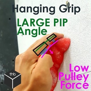
[Finger anatomy series 3/3] 3.The Proximal Interphalangeal joint or PIPNow that you know about the flexor tendons, the function of the pulleys and where they tend to pull, you should be able to guess why the PIP joint is so important!The PIP is the main joint that dictates the forces along the pulleys due to the position and angle of pull from the forces of the tendons.Demonstrated here is more of a hanging grip or an open handed grip. The angle of the PIP in this image is quite large and thus the resultant forces on the pulleys is minimal !! The SMALLER angle in the PIP = GREATER forces on the pulleys! Take this into consideration when thinking about how you grip your holds! I&;ll demonstrate with some more pictures in the following posts! posted on August 22, 2017 by Dr. Jonathan Leung | No comments by
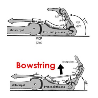
[Finger anatomy series 2/3] 2. Annular Pulleys These puppies prevent your finger from doing something known as bowstringing. These pulleys are fibrous bands that are used to maintain the contact of the finger tendon along the bone. What they do is transmit the force of the tendon into different angles of pull. Without them our hands wouldn&;t function very well and would look very weird. Bowstringing occurs when there has been a COMPLETE pulley rupture. With climbing the A2, A4 and A3 are most commonly injured!A2 is the largest of them all and attaches directly to bone. This one takes the brunt of the work when we go to climb or crimp!A3 attaches to whats known as the volar plate – it doesn&;t really attach directly to the bone! This is super super important when ADOLESCENTS or younger adults are climbing as an injury here can PERMANENTLY affect a GROWTH PLATE! It can potentially cause dramatic issues for their future. A rule of is if an adolescent complains of finger injury – SEEK PROFESSIONAL HELP. A4 attaches to the bone directly as well closer to the finger tip and is much smaller than the A2. How can you use this to help? Put less strain on your pulleys! Warm up with progressive bodyweight when crimping. Slow and steady wins the race. Feel free and content with any questions or concerns but up next is the PIP and how to crimp safer and stronger! posted on August 18, 2017 by Dr. Jonathan Leung | No comments by
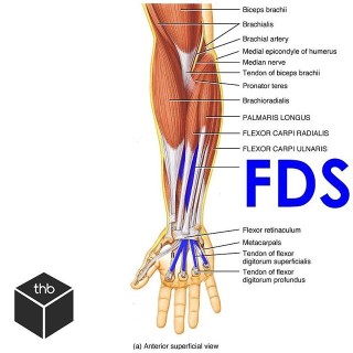
1. Flexor tendonsThe flexor tendons in the fingers occur because the muscles of the forearms become tendons as they enter the carpel tunnel into the hand.As you can see from the first image we have a bunch of other muscles of the forearm that affect motion of the hand and wrist. Well ignore those for now! First up is the Flexor digitorum superficialis or FDS. Swipe ️ and see it visualized again with its interaction with its deeper counterpart. The Flexor digitorum profundus or FDP is the deeper muscle that extends to the finger tip and helps us with flexing the entire fingerFDS is a tendon that splits into a V to allow the deeper FDP to run between it right to the tip! It inserts into the middle phalanx and flexes primarily the PIP joint. The design of these two tendons contribute to why we have such fine motor control at our fingers but it&;d be a shame if these tendons didn&;t have anything strong to hold them down! Next up. Pulleys. posted on August 17, 2017 by Dr. Jonathan Leung | No comments by
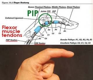
The finger is comprised of a number of joints, muscles, tendons, and many pulleys!Visualized here is your index finger! In the next posts we will will focus on 3 things: Flexor tendonsAnnular pulleysProximal Interphalangeal joint (PIP)There are many other components to the finger that are important to climbing. Injuries to these areas such as the joint capsules of each phalanx, each knuckle (capsule tissue injuries/capsulitis), the lumbrical muscles (another finger muscle injury source!) and the volar plates. (super super important for ADOLESCENT climbers! Attn: coaches) stay tuned. posted on August 17, 2017 by Dr. Jonathan Leung | No comments by
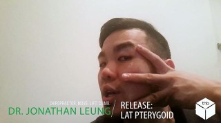
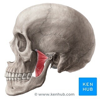
If you&;ve got ringing in the ears (tinnitus), headaches or neck pain that won&;t seem to go away even with treatment? Did you know your jaw could be the cause?.Pterygoid.The Pterodactyl&;s of your body – but no actually – these wing shaped muscles are notoriously hard to feel – especially extra-orally – they are the last 2 in the muscles of mastication and their function is to assist in chewing! (Again! big surprise!) Your pterygoids are comprised of 2 parts.Medial PterygoidsLateral PterygoidsThese muscles work together to help ELEVATE and PROTRUDE the jaw. The lateral pterygoid muscle is one to note because its the ONLY muscle of mastication that can actually help open the jaw.Not all muscles are meant to just simply be released! For example – if you have a nerve entrapment of the trigeminal nerve which is a nerve in your face which also controls some muscles of sound modulation (tensor tympani, tensor veli palatini) you can have RINGING in your ears or tinnitus. This may be due to too much INACTIVITY of muscles designed to hold the jaw in its proper forward position. If the mandible shifts too far back tension can be placed on the nerve causing some of your symptoms. Confused yet? For every joint in the body, there is an optimal position, often you do not want to simply jam the joint as far back as it can go – the same applies for the TMJ. Try out some of the stuff I&;ve posted but if you&;re really concerned just go get a screen done by a professional! Most will provide free consultations. Check yourself before you wreck yourself. series posted on May 4, 2017 by Dr. Jonathan Leung | No comments by
If you&;ve got ringing in the ears (tinnitus), headaches or neck pain that won&;t seem to go away even with treatment? Did you know your jaw could be the cause?.Pterygoids.The Pterodactyl&;s of your body – but no actually – these wing shaped muscles are notoriously hard to feel – especially extra-orally – they are the last 2 in the muscles of mastication and their function is to assist in chewing! (Again! big surprise!) Your pterygoids are comprised of 2 parts..Medial PterygoidsLateral Pterygoids.These muscles work together to help ELEVATE and PROTRUDE the jaw. The lateral pterygoid muscle is one to note because its the ONLY muscle of mastication that can actually help open the jaw..Got ringing in the ears or what&;s known as tinnitus? The lateral pterygoid may be the culprit. A hyperactive pterygoid has connections to the inner ear and tugging on this can create or cause that incessant ringing..Got a click in your jaw? The lateral pterygoid has a role in causing what is known as anterior disc displacement – or pulling that articular disc in your jaw forward slightly when hyperactive. Although this muscle may be a contributing factor – We have not even begun to go down the rabbit hole when talking about disc displacements. Those are best addressed on a case by case basis with a through assessment. series posted on May 2, 2017 by Dr. Jonathan Leung | No comments by
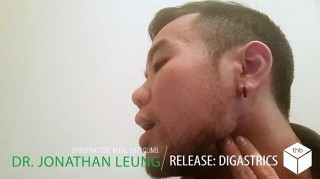
If you&;ve got headaches, neck pain or ringing in the ears that won&;t seem to go away even with treatment? Did you know your jaw could be the cause?.Digastrics. .Today we&;ll be going for the posterior belly of the digastric in 3 steps1. Gently perform some circular massage in the area under your ear and the angle of your mandible2. Put your finger behind the angle of the mandible and press forward toward the opening of the mouth or your eye3. Do this gently on both sides at once4. Take it slow and open your mouth or take deep breaths in and out5. Repeat 8-10xDisclaimer: this is a sensitive area so be gentle! series posted on April 21, 2017 by Dr. Jonathan Leung | No comments by
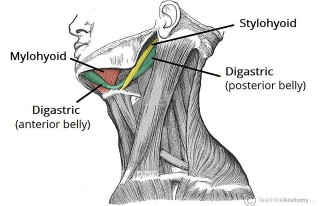
If you&;ve got headaches, tooth ache or neck pain that won&;t seem to go away even with treatment? Did you know your jaw could be the cause?.The Digastrics. .The Digastric muscles – by definition this double bellied muscle is a small muscle underneath the jaw – There are two bellies of the digastric: Posterior belly – attaches to the lower part of the skullAnterior belly – inner side of the mandible and the hyoid bone – a cartilaginous structure in your neckThis muscle helps you with swallowing and depressing the jaw. In patients with TMD, often the posterior belly of this muscle is tight.To muscle test it:.Put your fingers in the corner of your jaw and press forward and toward your eyes.If you swallow and feel this muscle pop – you&;re on the right spot.If you don&;t quite feel this one – don&;t worry. It&;s a smaller muscle which blends with a number of muscles into the front of the neck and is quite a tender point! Some practitioners themselves find difficulty muscle testing this on their patients. I just wanted to show you as much as I could for self treatment – This is number 3 of 4 so only one more!. Next up is the self release for it. series posted on April 20, 2017 by Dr. Jonathan Leung | No comments by
If you&;ve got ringing in the ears (tinnitus), headaches or neck pain that won&;t seem to go away even with treatment? Did you know your jaw could be the cause?——————–Now that you understand some of the basic movements of the jaw – we&;ll go into a little more detail about how the TMJ functions as a whole. The two smooth movements the TMJ have are:Rotation TranslationHere you will see how it works in motion – Jaw opening – During the first bit of jaw opening there is just pure rotation, and then as the jaw opens wider, the articular disc moves and there is a translation component. This video here demonstrates normal TMJ function. .Do you have a click? .If everything is moving well – you will have a smooth motion. If not, you may have a bit of a click or a pop and this is the articular disc getting displaced or pinned during this TRANSLATION phase of movement and then suddenly popping or snapping into place. Note that this processed is reversed on jaw closing and allows another opportunity for displacement or dysfunction to arise with the disc. . series posted on April 17, 2017 by Dr. Jonathan Leung | No comments by
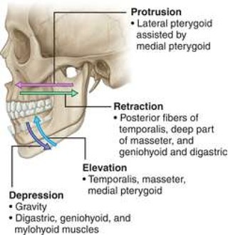
Ok. Back to basics. ——————–Now that you&;ve seen some of the self assessment of the jaw, we should understand the basic movements of the jaw. When both of your temporomandibular joints move in concert, you have the following 4 movements/terminologies:.Protrusion – jaw moves forwardRetraction – jaw pulls backwardElevation – jaw risesDepression – jaw drops .The above image shows you some of the muscles involved with each movement of the jaw..When the muscles of the TMJ are active on only one side you have the movement of Lateral deviation..Next up a video showing you the 2 main movements at the TMJ itself. Rotation and translation or spin and glide! . series posted on April 13, 2017 by Dr. Jonathan Leung | No comments by