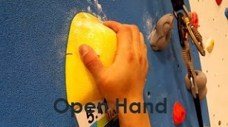
A little while ago I wrote up some details of every grip type. Finally getting around to posting them!OPEN HAND GRIPThis grip position places a lot less stress on the finger joints and tendons. The amount of force on your A2 pulley using the the crimp grip vs. the open hand grip is over 36x! So when you climb try your best to use this type of grip wherever you can.This position can be trained to become one of your strongest grip positions and is most effective on deep, rounded, sloper or pocket holds.Take home – Keep the angle between your distal and middle phalanx as open or large as possible! – Use the open hand grip EVERYWHERE! train with it, embrace it, become one with it. @hubclimbing posted on December 6, 2017 by Dr. Jonathan Leung | No comments by
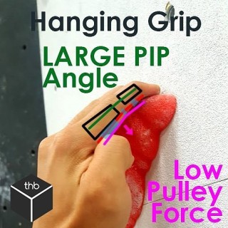
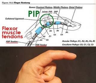
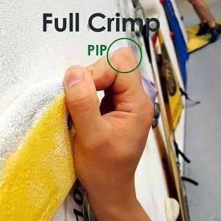
[Finger anatomy series 3/3] 3.The Proximal Interphalangeal joint or PIPNow that you know about the flexor tendons, the function of the pulleys and where they tend to pull, you should be able to guess why the PIP joint is so important!The PIP is the main joint that dictates the forces along the pulleys due to the position and angle of pull from the forces of the tendons.Demonstrated here is more of a hanging grip or an open handed grip. The angle of the PIP in this image is quite large and thus the resultant forces on the pulleys is minimal !! The SMALLER angle in the PIP = GREATER forces on the pulleys! Take this into consideration when thinking about how you grip your holds! I&;ll demonstrate with some more pictures in the following posts! posted on August 22, 2017 by Dr. Jonathan Leung | No comments by
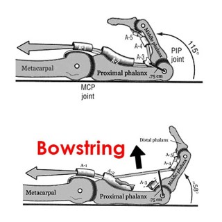
[Finger anatomy series 2/3] 2. Annular Pulleys These puppies prevent your finger from doing something known as bowstringing. These pulleys are fibrous bands that are used to maintain the contact of the finger tendon along the bone. What they do is transmit the force of the tendon into different angles of pull. Without them our hands wouldn&;t function very well and would look very weird. Bowstringing occurs when there has been a COMPLETE pulley rupture. With climbing the A2, A4 and A3 are most commonly injured!A2 is the largest of them all and attaches directly to bone. This one takes the brunt of the work when we go to climb or crimp!A3 attaches to whats known as the volar plate – it doesn&;t really attach directly to the bone! This is super super important when ADOLESCENTS or younger adults are climbing as an injury here can PERMANENTLY affect a GROWTH PLATE! It can potentially cause dramatic issues for their future. A rule of is if an adolescent complains of finger injury – SEEK PROFESSIONAL HELP. A4 attaches to the bone directly as well closer to the finger tip and is much smaller than the A2. How can you use this to help? Put less strain on your pulleys! Warm up with progressive bodyweight when crimping. Slow and steady wins the race. Feel free and content with any questions or concerns but up next is the PIP and how to crimp safer and stronger! posted on August 18, 2017 by Dr. Jonathan Leung | No comments by
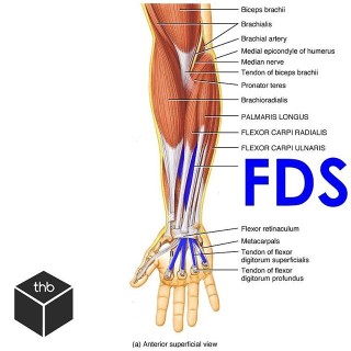
1. Flexor tendonsThe flexor tendons in the fingers occur because the muscles of the forearms become tendons as they enter the carpel tunnel into the hand.As you can see from the first image we have a bunch of other muscles of the forearm that affect motion of the hand and wrist. Well ignore those for now! First up is the Flexor digitorum superficialis or FDS. Swipe ️ and see it visualized again with its interaction with its deeper counterpart. The Flexor digitorum profundus or FDP is the deeper muscle that extends to the finger tip and helps us with flexing the entire fingerFDS is a tendon that splits into a V to allow the deeper FDP to run between it right to the tip! It inserts into the middle phalanx and flexes primarily the PIP joint. The design of these two tendons contribute to why we have such fine motor control at our fingers but it&;d be a shame if these tendons didn&;t have anything strong to hold them down! Next up. Pulleys. posted on August 17, 2017 by Dr. Jonathan Leung | No comments by
The finger is comprised of a number of joints, muscles, tendons, and many pulleys!Visualized here is your index finger! In the next posts we will will focus on 3 things: Flexor tendonsAnnular pulleysProximal Interphalangeal joint (PIP)There are many other components to the finger that are important to climbing. Injuries to these areas such as the joint capsules of each phalanx, each knuckle (capsule tissue injuries/capsulitis), the lumbrical muscles (another finger muscle injury source!) and the volar plates. (super super important for ADOLESCENT climbers! Attn: coaches) stay tuned. posted on August 17, 2017 by Dr. Jonathan Leung | No comments by
Swipe ️ to see all the climbing grip types. Climbers use their hands in a multitude of ways. This series of will demonstrate the various positions of climbing and grip types.Back to basics. Different hold positions.full crimphalf crimpopen crimppocketnarrow pinchfat pinchopen handPay attention to the positions of each grip and think about the angles between the MIDDLE knuckle in each hold type. This joint is the Proximal Interphalangeal joint or the PIP for short. Notice how the finger position in the pocket is very similar to a half crimp position? Sometimes even the open crimp/open hand position! Only on fewer fingers. The pinch grips are the SAME as the open grip position! Some of you advanced or pro climbers say Dr. Jon! That ain&;t all of them!… Well then here&;s a more exhaustive list but they all use the same concepts as above! What about slopers?Slopers use an open hand grip type to ensure maximum surface area contact for a friction based gripWhat about underclings? a jug hold with open hand/ half crimp grip stylesWhat of a Gaston? This along with the mantle is more of a pushing grip instead of pulling. Gaston is often in a half grip positionMantle is a open hand max surface area friction moveThe absolute best grip in terms of climbing longevity and strain on the fingers hand and pulleys is the OPEN HAND grip type. Remember this. Think about why it may be the case! My next post will showcase some of the anatomy of the finger focusing mainly on the pulleys! Stay tuned. posted on August 16, 2017 by Dr. Jonathan Leung | No comments by
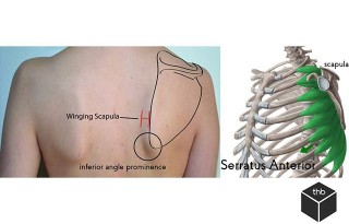
A Winging scapula is a result of weakness of the Serratus Anterior muscle or damage to the long thoracic nerve which supplies this muscle. The result is a protruding shoulder blade into the back.Test yourself – While wearing a tank top:Put yourself in a pushup position against the wallLean forward into your handsAsk somebody to take a picture for you.Do you notice a bump on one side versus the other?If you don&;t have a friend – go into the washroom and take a look at your shoulder heights.Do you notice one shoulder significantly lower than the other?If you do. You have scapular winging. This can affect your ability to lift, pull, or push objects. The reason is because scapular winging is a muscular imbalance which results in malpositioning of the shoulder blade. This disrupts the Scapulohumeral rhythm – which is the optimal ratio of movement when you lift your arm from your side above your head. If this is a result from a long thoracic nn entrapment Active Release Therapy or ART can definitely help with this nervous entrapment. Proper positioning and movement of the scapula is critical for full and normal shoulder range of motion. Without it, your scapular is SICK! (Scapular malposition, Inferior medial border prominence, Coracoid pain and malposition, and dysKinesis of scapular movement) SICK scapula or scapular dyskinesis refers to an injury resulting from overuse and fatigue of the muscles that stabilize and provide motion for the scapula.To counteract it we want to:. Treat the underlying soft tissue adhesionsStrengthen the affected musclesGrease the groove and provide stimuli to maintain the positioning and movement of the scapula posted on May 30, 2017 by Dr. Jonathan Leung | No comments by
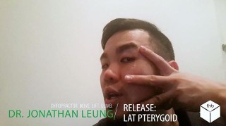
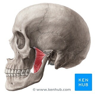
If you&;ve got ringing in the ears (tinnitus), headaches or neck pain that won&;t seem to go away even with treatment? Did you know your jaw could be the cause?.Pterygoid.The Pterodactyl&;s of your body – but no actually – these wing shaped muscles are notoriously hard to feel – especially extra-orally – they are the last 2 in the muscles of mastication and their function is to assist in chewing! (Again! big surprise!) Your pterygoids are comprised of 2 parts.Medial PterygoidsLateral PterygoidsThese muscles work together to help ELEVATE and PROTRUDE the jaw. The lateral pterygoid muscle is one to note because its the ONLY muscle of mastication that can actually help open the jaw.Not all muscles are meant to just simply be released! For example – if you have a nerve entrapment of the trigeminal nerve which is a nerve in your face which also controls some muscles of sound modulation (tensor tympani, tensor veli palatini) you can have RINGING in your ears or tinnitus. This may be due to too much INACTIVITY of muscles designed to hold the jaw in its proper forward position. If the mandible shifts too far back tension can be placed on the nerve causing some of your symptoms. Confused yet? For every joint in the body, there is an optimal position, often you do not want to simply jam the joint as far back as it can go – the same applies for the TMJ. Try out some of the stuff I&;ve posted but if you&;re really concerned just go get a screen done by a professional! Most will provide free consultations. Check yourself before you wreck yourself. series posted on May 4, 2017 by Dr. Jonathan Leung | No comments by
If you&;ve got ringing in the ears (tinnitus), headaches or neck pain that won&;t seem to go away even with treatment? Did you know your jaw could be the cause?.Pterygoids.The Pterodactyl&;s of your body – but no actually – these wing shaped muscles are notoriously hard to feel – especially extra-orally – they are the last 2 in the muscles of mastication and their function is to assist in chewing! (Again! big surprise!) Your pterygoids are comprised of 2 parts..Medial PterygoidsLateral Pterygoids.These muscles work together to help ELEVATE and PROTRUDE the jaw. The lateral pterygoid muscle is one to note because its the ONLY muscle of mastication that can actually help open the jaw..Got ringing in the ears or what&;s known as tinnitus? The lateral pterygoid may be the culprit. A hyperactive pterygoid has connections to the inner ear and tugging on this can create or cause that incessant ringing..Got a click in your jaw? The lateral pterygoid has a role in causing what is known as anterior disc displacement – or pulling that articular disc in your jaw forward slightly when hyperactive. Although this muscle may be a contributing factor – We have not even begun to go down the rabbit hole when talking about disc displacements. Those are best addressed on a case by case basis with a through assessment. series posted on May 2, 2017 by Dr. Jonathan Leung | No comments by
&;Creamsicle toss&; this covered in the chalk and blood of my brothers before me.Beta tip:If you don&;t have the accuracy or desire to throw to a pocket/crimp (that you cant see because the red hides it!) use momentum and power to skip right past it. The next hold is a jug. I&;d rather throw an extra 4 inches than smash my hand a hundred times.Maximum effort climb but as fun as it gets!Thanks for the support and angles @heyyitscheryl Chris! Leen! Rudy! ction posted on April 29, 2017 by Dr. Jonathan Leung | No comments by